Physio Blogfrom the team at South Coast Physiotherapy

James Gasper
New Physio Starting
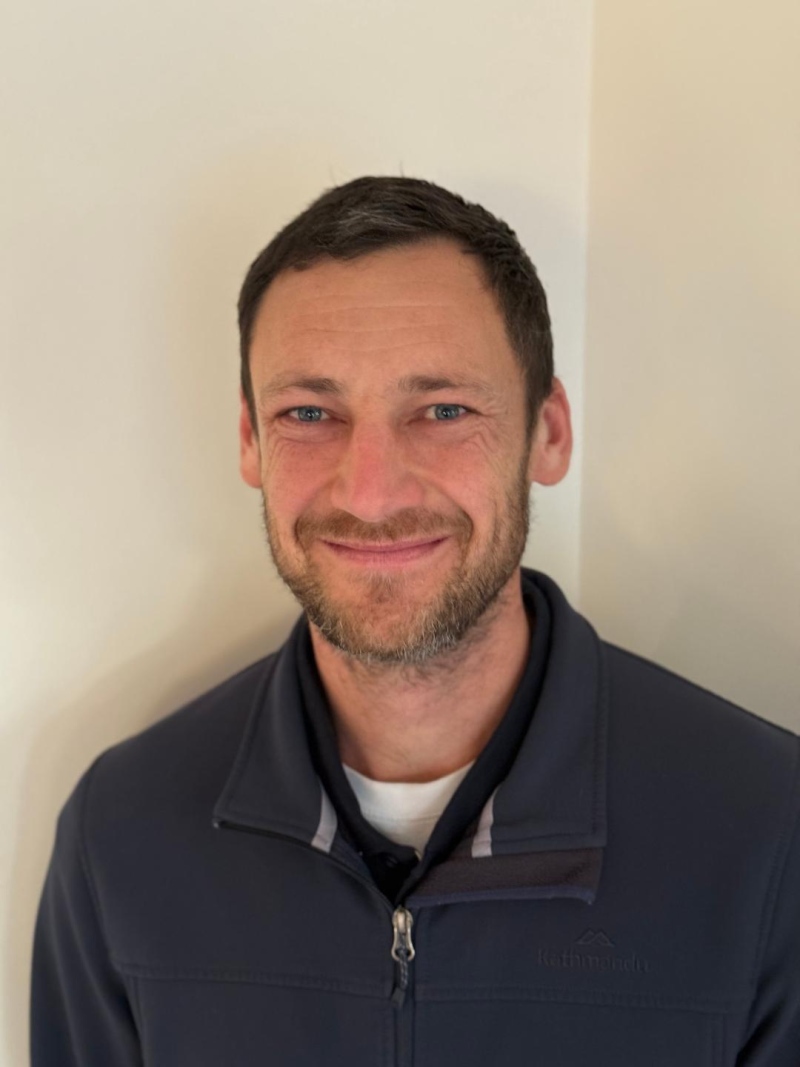
We are delighted to welcome Matthew Ott to the South Coast family.
Matthew is a highly experienced physiotherapist who has recently moved down to the Mornington Peninsula from NSW. He is currently open to see patients on Tuesdays and Wednesdays.
Understanding the Vestibular System and how Physiotherapy can help with Vertigo
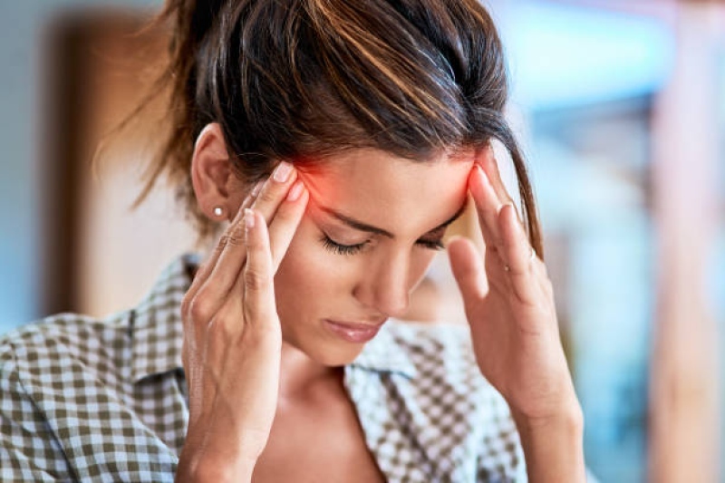
The vestibular system helps us control balance, eye movements, and our sense of position in space. The peripheral vestibular apparatus is located in the inner ear and consists of the bony labyrinth—made up of three semicircular canals, the utricle, and the saccule. Conditions affecting this part of the system are referred to as peripheral vestibular disorders.
When the vestibular system is disrupted, people may experience vertigo—commonly described by patients as dizziness, spinning, swaying, or disequilibrium.
Common Peripheral Vestibular Conditions
There are many medical causes of dizziness, so it is important that a medical evaluation by a GP, neurologist, or ENT specialist is undertaken. Some of the most common vestibular-related conditions include:
- Benign Paroxysmal Positional Vertigo (BPPV)
Caused by dislodged calcium carbonate crystals (otoconia) moving into the semicircular canals. This leads to brief episodes of vertigo triggered by changes in head position such as rolling in bed or looking up. - Menière’s Disease
Thought to result from an abnormal build-up of endolymphatic fluid in the inner ear, causing episodes of vertigo, fluctuating hearing loss, tinnitus, and a sensation of fullness in the ear. - Labyrinthitis
Inflammation of the entire inner ear (labyrinth), usually viral, affecting both balance and hearing. It typically presents with acute vertigo, nausea, and hearing loss. - Vestibular Neuritis
Inflammation of the vestibular nerve, usually viral, impairing balance but not hearing. It is characterised by sudden onset of severe vertigo, nausea, and unsteadiness that may last for days.
Physiotherapy and Vestibular Care
Physiotherapists who have completed additional vestibular training play a vital role in the assessment and management of peripheral vestibular conditions.
The most common cause of vertigo seen in physiotherapy practice is BPPV.
BPPV and the Modified Epley Manoeuvre
BPPV is typically diagnosed with a positional test (e.g. Dix-Hallpike), in which vertigo is reproduced and nystagmus (involuntary, repetitive eye movement) is observed. The direction, duration, and type of nystagmus—along with the head position—help determine which canal is affected.
Treatment usually involves a Canalith Repositioning Manoeuvre (CRM) (e.g. Modified Epley) which involves moving the head in particular position sequentially to move the otoconia (crystals) into the correct position. These choice of which manoeuvres is used to treat the BPPV is dependent on which canal is affected. Success rates for CRM for BPPV have been shown to be as high as 85% after one treatment and up to 95% resolution within 1–3 sessions. There is minimal risk of recurrence or complications when performed correctly
Vestibular Rehabilitation for Other Conditions
Other vestibular conditions (e.g. Menière’s disease, vestibular neuritis, labyrinthitis) may lead to vestibular hypofunction—where the sensory input from one or both vestibular apparatus is impaired, resulting in reduced balance function and dizziness.
In these cases, physiotherapists use Vestibular Rehabilitation Therapy (VRT) to help the brain compensate, habituate, and adapt to altered sensory input. This typically includes:
- Balance retraining
- Gaze stabilisation exercises (to retrain the vestibulo-ocular reflex)
- Habituation exercises (to reduce motion sensitivity)
- Graded exposure to movement and visual complexit
Systematic reviews and clinical guidelines show moderate to strong evidence supporting VRT for improving balance, reducing dizziness, and restoring function in vestibular hypofunction.
Conclusion
Peripheral vestibular conditions are a common cause of vertigo and dizziness. Many are highly treatable, sometimes with near-complete symptom resolution. With accurate diagnosis and evidence-based physiotherapy, patients often experience significant improvement in both symptoms and quality of life.
If you are experiencing persistent dizziness or balance issues, consider booking an assessment with a physiotherapist trained in vestibular rehabilitation.
References
- Neuhauser, H. K. et al. (2005). The epidemiology of dizziness and vertigo. Journal of Neurology, Neurosurgery & Psychiatry, 76(5), 710–715.
- Bösner, S. et al. (2018). Dizziness in primary care: diagnostic accuracy of clinical tests. BMC Primary Care, 19(1), 33.
- Agrawal, Y. et al. (2009). Vestibular dysfunction in older individuals. Archives of Internal Medicine, 169(10), 938–944.
- Hilton, M. P., & Pinder, D. K. (2014). The Epley manoeuvre for posterior canal BPPV. Cochrane Database of Systematic Reviews, (12).
- Li, J. C. et al. (2023). Modified vs standard Epley manoeuvre: RCT. Frontiers in Neurology, 14, 1328896.
- McDonnell, M. N. et al. (2015). Vestibular rehabilitation for unilateral vestibular hypofunction: Cochrane Review. Cochrane Database of Systematic Reviews, (1).
New Audologist

South Coast Physiotherapy welcomes Audiology Services
We are excited to announce the expansion of our services to include professional
Audiological care at our clinic. Whether you are looking for a hearing assessment, hearing
aid fitting, wax management, or tinnitus support our highly qualified Audiologist is here to
guide you towards better hearing health.
Phung La is an experienced Audiologist with over 15 years of expertise in hearing
healthcare. Throughout her career, Phung has been dedicated to helping patients
overcome their hearing challenges, enhance their quality of life and reconnect with loved
ones through personalized treatment plans tailored to individual needs.
Graduating from the University of Melbourne’s Master of Clinical Audiology program in
2009, Phung has worked in a diverse range of roles specializing in adult hearing
rehabilitation, Paediatrics, Workcover and Complex Adults.
Phung has also worked for many years as a Clinical Specialist (and later Sales Director) for
a leading hearing aid manufacturer, so she is well versed in fitting the latest and most
advanced hearing solutions.
Phung is truly passionate about making a positive impact on the lives of people with
hearing impairment, improving their self-confidence and ensuring they don’t miss out on
life’s precious moments.
To make an appointment please call 0481335432 or visit hear4life.com.au
Focus Areas:
- Comprehensive Hearing assessments for adults and children over 4 years.
- Government Hearing Services Program for Pensioners
- Ear cleaning and Ear Wax removal
- Tinnitus management and counselling
- Hearing aid fitting and rehabilitation
- Workcover assessment and hearing device fitting
- DVA (Veterans) assessment and hearing device fitting
- Pre-Employment Hearing checks
Achilles Tendinopathy

The Achilles tendon is one of the most commonly injured tendons in the body and unsurprisingly it is therefore an injury we see a lot of. This blog will be a quick run down on the fundamentals of what an Achilles tendinopathy is and how we treat them.
What is a tendinopathy?
Unlike an acute muscle tear or a broken bone a tendinopathy is a more complex multifaceted injury. It is generally an overuse injury where the tendon (tendons are what attach our muscles to our bones) has been repeatedly pushed beyond its capacity. These injuries are characterized by pain, a decline in function and reduced exercise tolerance. Structurally we see a disruption of the collagen fibres and a deregulation of the extracellular matrix – the building blocks of the tendon and why we see the reduced function. There is also increased inflammatory mediators and sensory nerve innervation – which explains the pain.
Achilles Tendon
We have tendons all over our body and these injuries are seen in the shoulder with rotator cuff tendons, the knee with the patella tendon and the hip with the gluteal tendons (as well as the elbow, wrist and groin). The Achilles tendon (named after the famous Greek Warrior God) is the largest and strongest tendon in the human body. It is approximately 15cm in length and rotates clockwise up to 90 degrees, running from the soleus and gastrocnemius muscles and inserting into the back of the heel. The tendon is surrounded by a paratenon allowing it to glide back and forth. It is made up of Type I collagen giving it the ability store and release energy so that it can stretch and recoil like a spring in response to the stretch/shortening cycle of the calf complex – an important action for locomotion.
This unique structure allows the Achilles to withstand fast and dynamic loads, which can be up to 7 x a persons body weight during running and jumping activities.
It also makes rehabilitation of this tendon very challenging.
Loading Programs
As with all tendinopathies the key to successful management is load control. This goes back to the original cause of the injury that, in the majority of cases, is due to the tendon being subjected to excessive loads for too long.
In severe cases, for the Achilles tendon, this can result in needing a Cam boot to completely unload the tendon. In more moderate cases, a simple heel wedge in a supportive pair of shoes is adequate.
Loading exercises are then prescribed at different intensities depending on the stage of the tendinopathy. These exercises will range from static holds (isometrics) in the initial stages, slow heavy resisted movements in the mid-stages and fast resisted movements (plyometrics) in the later stages.
For the Achilles tendon, in severe cases, an initial exercise may be a simple seated calf raise with limited loading through the tendon. This would then be gradually progressed over a number of months. End stage can involve hopping drills and single leg loads of up to 1.5 x body weight.
Throughout this program the alignment of the foot and ankle need to be monitored to ensure efficient load transfer through joints and tendons. In some cases the exercises have to be modified to the individual. An example of this would be when the patient has an arthritic great toe – this will alter their ability to perform a calf raise.
Return to Running
As we work through this loading program our rehab becomes more functional and we try and make it specific to the individuals goals.
A good example of this is getting athletes back running – with Achilles tendinopathies being a frequent injury associated with both competitive and recreational runners. A safe return to running program is an important part of their rehab process. The loads that go through the tendon when running are at a high tempo and can be between 5-8 x body weight. It is therefore wise to introduce running gradually with slow exposure to these loads ensuring that the tendon has time to adapt.
Take home message
- The Achilles tendon is a complex structure capable of tolerating impressive loads that enables us to walk, run and jump.
- An Achilles tendinopathy is when this tendon has been loaded beyond is capacity causing a number of pathological changes.
- To manage these injuries a loading program needs to be introduced – but getting the right intensity of loads with the right timeframes can be frustrating and is likely to be different for each individual.
- This program needs to be progressed through stages and in line with the individuals goals – not all rehab programs will be at the same intensity.
New Remedial Massage Therapist

We are excited to introduce, Tegan, our new Remedial Massage Therapist.
Tegan holds a diploma in Remedial Massage and has been running her own massage business in Flinders for the past 2 years.
She will be available in rooms on Mondays and will be a brilliant adjunct in the treatment and ongoing management of joint and muscle injuries as well helping in injury prevention.
NDIS Update from Raquel Poyser

Over the past twelve months, South Coast Physiotherapy have continued to provide Physiotherapy Services for our valued NDIS clients who now total just under 100.
These services range from providing hands on therapy to advice regarding assistive technology eg, wheelchairs, Ebikes and trikes, blue tooth operated hi lo beds to supporting participants with on land or in pool exercise programs to training for support workers.
Our Physiotherapists work and provide services for our NDIS clients in a variety of settings depending on what suits our clients and their situation best. These places of work may include our clinic rooms in Tootgarook, in a participant’s own home, supported accommodations, Adult Day Programs and in the community eg. YAWA, PARC.
South Coast Physiotherapy clinicians travel far and wide to assist our NDIS clients including Rye, Hastings, Cranbourne and Carrum and locations in between.
Sometimes our Physiotherapists are required to travel further up the train line to places like Mentone if we are meeting with a client at a provider to review and make modifications to their wheelchair.
Our therapists regularly work closely with large therapy and support teams supporting our NDIS clients to optimise their functional outcomes and assist them to work towards their NDIS goals.
At South Coast Physiotherapy we take pride in our work helping improve the lives of people living with a disability and striving to improve the services we offer our valued NDIS clients.
We look forward to supporting more people achieve great things in 2024!
Raquel Poyser
Neurological Physiotherapist
Christmas Opening Hours
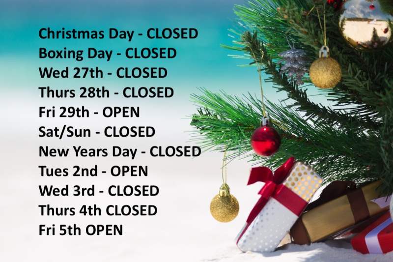
From everyone at South Coast Physiotherapy we wish you all a very Merry Christmas and Happy New Year.
We will have limited opening hours over this period but will be back to full hours on 8th January.
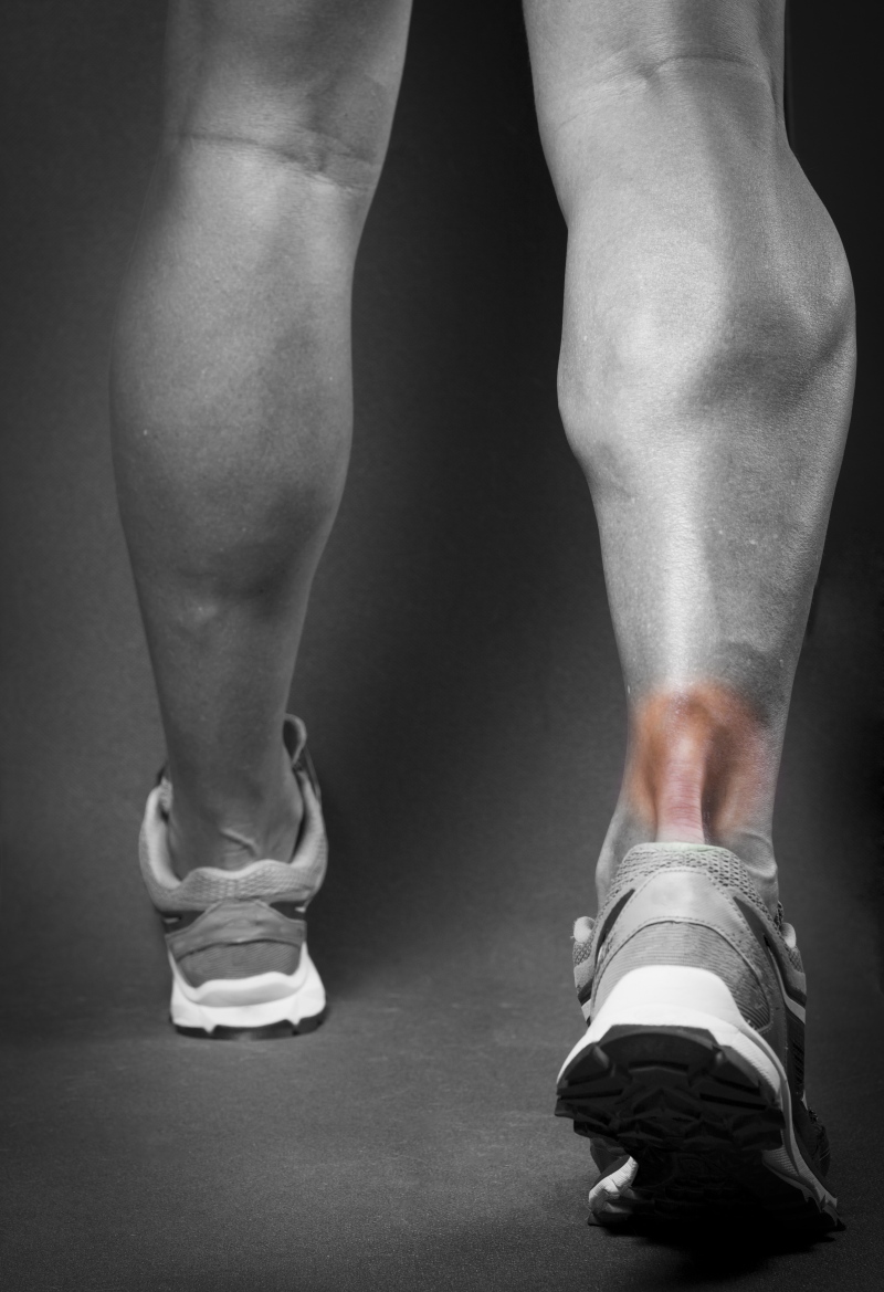
Calf Injuries

From Sam Kerr to Nathan Lyon to Will Skelton - calf injuries plagued our Australian sports stars through 2023.
Injuries to this muscle complex are in fact a very common sporting injury with only hamstrings and quadriceps being more frequently injured in sporting populations.
The superficial calf complex is known as the triceps surae and comprises of 3 muscles – the gastrocnemius, soleus and plantaris. Interestingly it has been found that injuries to the individual tricep surae muscles can be sport specific with American Football players mostly injuring the gastrocnemius and Australian Rules Footballers mostly injuring the soleus (1,2)
A gastrocnemius strain will occur with a sudden ballistic movement with the knee in full extension. This is because the muscle attaches above the knee joint and is therefore exposed when the knee is fully extended. The injury is more prone in the medial head of the muscle – due to this part of the muscle experiencing more loads.
A soleus strain is often associated as an overuse injury. This muscle attaches below the knee and is therefore loaded with the knee in flexion – although it is also loaded with knee extension.
On examination these tears can have a palpable defect at the site of the tear if they are high grade. They will have pain when stretching the calf and on resisted plantar flexion movements such as performing a heel raise.
These tears are categorized based on the degree of structural damage. Recently, with MRIs being more readily available, this process has become more specific which has allowed medical teams a more precise prediction of outcomes. One example of this is the British Athletic Classification, which is used globally, and groups muscle injuries based on the location of injury and the involvement of different soft tissue structures – along with the degree of structural damage. This is how the medical teams new that Nathan Lyon’s Ashes tour was over but that Sam Kerr had a chance of making the latter stages of the world cup – (Will Skelton new his tournament was over – but this was more due to the Wallabies form than his injury!!).
The rehabilitation of these injuries is split into different phases – with the initial phases focusing on protection and gradual restoration of ankle movement to the final stages of returning to running, jumping and ultimately sport.
Although a clinician can give an estimation of how long it will take an individual to progress through these phases based on the classification of the injury it is important that the individual meets set criteria before moving through the phases. For example, for a calf injury, being able to complete single leg heel raises is important before starting a return to running program in order to minimize the re-injury risk.
Once the patient has returned to their full capacity it is important that they maintain good strength and mobility through their calf complex in order to minimize the risk of future injury. This will often mean continuing with some of the later stage rehabilitation exercises and incorporating them into their training schedule.
Achilles Tendon Rupture - The importance of rehabilitation
Named after the famous Greek Warrior God the Achilles tendon is the largest and strongest tendon in the human body. It is approximately 15cm in length and rotates clockwise up to 90 degrees, running from the soleus and gastrocnemius muscles and inserting into the back of the heel. The tendon is surrounded by a paratenon allowing it to glide back and forth. It is made up of Type I collagen giving it the ability store and release energy so that it can stretch and recoil like a spring in response to the stretch/shortening cycle of the gastrocnemius and soleus muscles – an important action for locomotion.
This unique structure allows the Achilles to withstand fast and dynamic loads, which can be up to 7 x a persons body weight during running and jumping activities (1). It also makes rehabilitation of this tendon very challenging.
It is the most commonly injured tendon in the human body and complete ruptures are reported from 11 to 37 per 100,000 population and are currently on the increase (1).
A ruptured tendon is when there is a complete full thickness tear through the tendon fibres.
An Achilles rupture will often be a traumatic injury with acute pain and a sudden inability to walk. Patients describe a popping sensation at their heel and a feeling of being kicked.
For the Achilles this injury has a bimodal age distribution with it occurring in 2 groups of the population – those between 25-40yrs and those over 60 yrs (2). It is generally an acute injury in the younger group and can be more of a chronic presentation in the older population – although as our aging population strive to remain active we are seeing an increasing number of acute ruptures in this older demographic.
This bimodal age distribution is significant when it comes to deciding on the best management with rehabilitation being governed by the age and health of the tendon. As a physiotherapist rehabilitating an Achilles rupture – both when surgically or non-surgically managed - my rehabilitation program and my expected outcomes would be different for a 25 year old compared to a 65 year old.
Unsurprisingly a rupture to this tendon is a debilitating injury and rehabilitation is on par with an ACL reconstruction of the knee.
Management of these injuries have always been somewhat controversial with surgical management being the traditional choice due to the high re-rupture rates and the risks of tendon lengthening in conservatively managed patients (3). However the risk of complications with surgery – including nerve damage, wound infection and scar adhesions - have lead to the pursuit of improved non-surgical management.
Over the last 20 years there has been a development of stricter rehabilitation protocols aimed at restoring tendon function. In particular the introduction of early weight bearing - this has been shown to promote the synthesis of type I collagen which is vital in restoring the tendons load resistant properties mentioned earlier (4). When these developments, which have also included functional orthosis and graded angles of plantar flexion, are introduced into non-surgical management protocols they have lead to a reduction in the non-surgical re-rupture rates and an improvement in long term function resulting in higher a return to work and to recreational activities (5,6).
We are now in a position where non operative management can be recommended for a large proportion of Achilles ruptures knowing that the outcomes will be similar to surgical repairs without the risks of surgery - as long as rehabilitation protocols are followed (7,8).
The development of rehabilitation protocols have assisted both surgically and non-surgically managed Achilles ruptures. These protocols are aimed at stimulating tendon healing and restoring the mechanical properties through a graded program of exercises and activities. These programs will differ from one patient to the next due to age, general health and patient goals – it is therefore beneficial to have an experienced clinician monitoring and progressing these protocols.
Although an Achilles rupture is still a serious injury, which requires a long period of rehabilitation, whether treated surgically or non-surgically, the development of rehabilitation protocols over the last 20 years has meant we are now able to restore the tendon back to withstanding fast and dynamic loads meaning more people are returning to their work and recreational activities.
- Jarvinen T A H et al. Achilles tendon disorders: Etiology and epidemiology. Foot Ankle Clin. 2005
- Moller A et al. Increasing incidence of Achilles tendon rupture. Act Orthop Scand. 1996
- Waterston S, Maffulli N, Ewen W. Subcutaneous rupture of the Achilles tendon: basic science and some aspects of clinical practice. Be J Sports Med 1997; 31:285-298
- Valkering KP et al. Functional weight-bearing mobilization after Achilles tendon rupture enhances early healing response: a single blinded randomized controlled trial. Knee Surg Traumatol Arthosc. 2017.
- Soroceanu A, Sidhwa F, Aarabi S, et al. Surgical versus nonsurgical treatment of acute achilles tendon rupture. J Bone Joint Surg Am 2012;94:2136–43.
- Winson D, MacNair R, Hutchinson A, Owen N, Evans R, Williams P. Delayed Achilles tendon rupture presentation: Non-opertaive management may be the SMART choice. The Foot 2020.101724
- Willits K, Amendola A, Bryant D, et al. Operative versus nonoperative treatment of acute Achilles tendon ruptures: a multicenter randomized trial using accelerated functional rehabilitation. J Bone Joint Surg Am 2010;92:2767–75.
- Wallace R, Heyes G, Michael A. The non-operative functional management of patients with a rupture of the tendo Achillis leads to low rates of re-ruputre. J Bone Joint Surg Br 2011;93-B:1362-6
Merry Christmas
We are having a Christmas break but will back fresh and ready to go for the New Year - Merry Christmas